Breadcrumb
Uncommon Presentation of a Residual Cyst
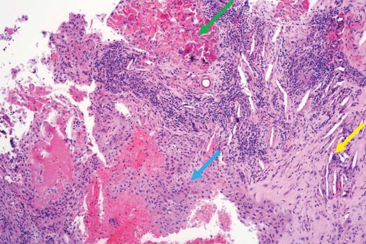
The histologic examination shows an inflamed and distorted cyst at 40× magnification. The green arrow indicates the presence of Rushton bodies, the yellow arrow points to cholesterol clefts with multinucleated giant cells, and the blue arrow highlights an inflamed cystic lining with neutrophil exocytosis.
What is it?
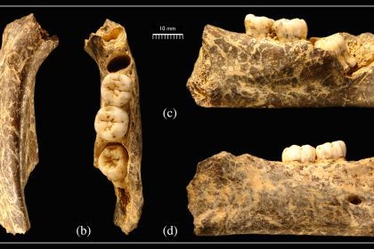
This is a case study of an 82-year-old male patient with jaw pain, which turns out to be a rare case where an X-ray shows a residual cyst with unusual calcium deposits inside. Cysts are abnormal hollow spaces within the bone, often lined by a thin layer of cells. These cells can come from tooth-related tissues (odontogenic) or other sources (non-odontogenic). Residual cysts are a type of inflammatory cyst that forms when parts of an earlier cyst are left behind after treatment. These often originate from untreated radicular cysts and occur in areas where teeth are missing. They are usually painless and found accidentally during dental X-rays. The lead author on the case study was a DDS student at the Dugoni School.
What problem does it aim to solve?
Cysts can be symptomless or cause mouth pain; treating them successfully requires accurate diagnosis not just of the cyst’s existence, but of its type. It’s important to develop, document and share best practices.
How does it work?
“A multidisciplinary approach involving oral surgery, oral and maxillofacial radiology, and oral pathology specialists was essential in managing this case. This collaborative effort ensured a holistic view of the patient’s dental condition and thus, a more personalized differential diagnosis.”
What are the real-world implications?
“This case underscores the importance of considering a cyst with dystrophic calcifications in the differential diagnosis of unilocular radiolucent lesions with radiopaque internal entities. Comprehensive patient assessment and a multidisciplinary treatment approach are critical for effective management and prevention of recurrence in such complex cases.”
What are the next steps?
This report emphasizes that when diagnosing unusual bone lesions with both dark and light areas on X-rays, dentists should consider residual cysts with calcifications as a possibility. It also highlights the importance of evaluating the overall dental condition to make an accurate diagnosis.
Source
Zamanian, N., Karimi, A., Bukhari, J., Davies, A. J., Mashkoor, F., & Daly, L. B. (2024). Uncommon Presentation of a Residual Cyst. Journal of the California Dental Association, 52(1). https://doi.org/10.1080/19424396.2024.2413577
Authors
Nazgol Zamanian
International Dental Studies program Class of 2025, University of the Pacific Arthur A. Dugoni, School of Dentistry
A. Karimi
International Dental Studies program Class of 2026, University of the Pacific, Arthur A. Dugoni School of Dentistry
J. Bukhari
BeamReaders, New York, USA
A. J. Davies
Department of Oral and Maxillofacial Surgery, University of the Pacific, Arthur A. Dugoni School of Dentistry
F. Mashkoor
Department of Oral and Maxillofacial Surgery, University of the Pacific, Arthur A. Dugoni School of Dentistry
L. B. Daly
Department of Diagnostic Sciences, University of the Pacific, Arthur A. Dugoni School of Dentistry